Treating diabetic retinopathy
Treating diabetic retinopathy is a critical aspect of preserving vision and maintaining the overall quality of life for patients suffering from diabetes. Diabetic retinopathy is a common complication of diabetes that affects the blood vessels in the retina – the light-sensitive tissue at the back of the eye.
According to the National Eye Institute, approximately 7.7 million Americans are estimated to have diabetic retinopathy, and that number is expected to rise to 14.6 million by 2050. Early detection and intervention can significantly reduce the risk of vision loss, making it vital for patients with diabetes to undergo regular eye exams and follow a personalized treatment plan.
The Role of The Optometrist and Ophthalmologist in the Management of Diabetic Retinopathy
The role of the optometrist and ophthalmologist in the management of diabetic retinopathy is distinct yet synergistic. Optometrists, as primary eye care professionals, are often the first line of defense in detecting diabetic retinopathy. They conduct comprehensive eye examinations and utilize advanced diagnostic technologies to identify early signs of the condition. Once detected, optometrists play a crucial role in patient education, explaining the implications of the disease, and offering advice on lifestyle adjustments to mitigate its progression. Ophthalmologists, on the other hand, are medical doctors specialized in eye and vision care who have completed additional training to handle more complex cases. In the context of diabetic retinopathy, they are often involved when the condition requires more invasive treatments such as laser surgery, vitrectomy, or injection of medications into the eye. Their expertise is vital in managing advanced stages of the disease and mitigating complications. Together, these professionals form a united front in the comprehensive management of diabetic retinopathy, combining preventative and therapeutic measures for optimal patient care.
Treatment Options for Diabetic Retinopathy
There are several treatment options available for diabetic retinopathy, which can vary depending on the stage of the condition and the specific needs of each patient. These treatments are designed to slow down or halt the progression of retinopathy, minimize the risk of vision loss, and, in some cases, improve visual acuity. Key treatment options for diabetic retinopathy include:
Blood sugar management
The cornerstone of any treatment plan for diabetic retinopathy is effectively managing blood sugar levels. Patients with well-controlled diabetes are less likely to experience vision-threatening complications, so it's essential to work closely with your healthcare team to achieve optimal blood sugar control.
Blood pressure and cholesterol control
High blood pressure and elevated cholesterol levels can contribute to the progression of diabetic retinopathy. Maintaining optimal blood pressure and cholesterol levels through medication and lifestyle changes is a vital aspect of managing the condition.
Laser treatment
Laser photocoagulation is a standard treatment option for diabetic retinopathy, especially for the advanced stage known as proliferative diabetic retinopathy (PDR). The laser seals off leaking blood vessels and discourages the growth of new, abnormal blood vessels that can lead to vision loss.
Anti-VEGF injections
Vascular endothelial growth factor (VEGF) is a protein that promotes the growth of new blood vessels. In some cases of diabetic retinopathy, anti-VEGF medications can be injected directly into the eye to inhibit the growth of abnormal blood vessels and decrease fluid leakage.
Vitrectomy
In advanced cases of diabetic retinopathy, a surgical procedure called vitrectomy may be necessary. During a vitrectomy, the vitreous gel is removed from the eye to clear blood and scar tissue, allowing for better light transmission to the retina.
Corticosteroid injections
In some instances, corticosteroid injections can help reduce inflammation and alleviate symptoms of diabetic macular edema – a common complication of diabetic retinopathy that can lead to central vision loss.
As your local optometrist, [mbv name="token-primary-doc-lastname"] is committed to providing personalized care tailored to your unique needs. Our approach to diabetic retinopathy involves a comprehensive evaluation including high definition imaging, a detailed discussion of your treatment options, and close collaboration with your primary care physician or endocrinologist to ensure the best possible outcomes. Through a combination of state-of-the-art technology and compassionate care, our team is dedicated to preserving your vision and helping you maintain a high quality of life.
Act Now to Protect Your Vision
Don't wait until it's too late – early detection and intervention are crucial in managing diabetic retinopathy and preventing vision loss. Schedule an appointment with [mbv name="practice-name"] in [mbv name="token-practice-city"] today to discuss your eye health, review your treatment options, and develop a customized plan that addresses your specific needs. Together, under the guidance of our eye care professional, [mbv name="token-primary-doc-lastname"], we can work towards preserving your vision and enhancing your overall well-being. Call us at [mbv name="token-practice-phone"] or [mbv name="token-appointment-link-with-text"] to secure your appointment. We look forward to helping you maintain optimal eye health and empowering you to take control.
Patients with diabetic retinopathy visit our clinic from all over [mbv name="practice-state-full"], and we are proud to be a leading provider of medical eye care services for patients from [mbv name="token-practice-city"], [mbv name="token-location-secondary1"], [mbv name="token-location-secondary2"], and [mbv name="token-location-secondary3"].
Read More
Is foggy vision troubling you? Let our experienced optometrist restore your sight – book an appointment today and see the difference!
The term foggy vision often refers to a visual disturbance characterized by blurred, cloudy, or hazy vision, which can make seeing clearly challenging. As your local optometrist, we recognize the significance of maintaining optimal eye health and are committed to offering our patients the latest and most accurate information on a variety of eye conditions, including this particular issue. In this blog, we will explore the causes, symptoms, and treatments for this vision problem, while also sharing important statistics about its prevalence.
Take Our Online Double Vision Assessment
Is blurry, fuzzy, or double vision impacting your quality of life and vision? Take our online double vision assessment to help identify if you may have an underlying vision problem that is causing diplopia (double vision) /neuro/double-vision-quiz/
Causes of Foggy Vision
There are numerous potential causes of foggy vision, including refractive errors, cataracts, glaucoma, age-related macular degeneration (AMD), dry eyes, corneal diseases, and even certain medications.
- Refractive errors, such as myopia (nearsightedness), hyperopia (farsightedness), presbyopia (age related farsightedness), and astigmatism, are the most common causes, affecting approximately 60% of the global population. Learn more about nearsightedness and farsightedness here (/ocular-disease/understanding-nearsightedness-and-farsightedness-causes-symptoms-and-diagnosis/).
- Cataracts, a clouding of the lens, is another leading cause, impacting nearly 24.4 million Americans aged 40 and above. Learn more about cataracts here (/ocular-disease/cataracts/).
- Glaucoma, a group of eye conditions characterized by damage to the optic nerve, can also result in foggy vision, with over 3 million Americans affected. AMD, which affects the central part of the retina, is a leading cause of vision loss in people aged 50 and older, with around 11 million cases in the United States alone. Learn more about AMD here (/low-vision/age-related-macular-degeneration-and-low-vision/).
- Dry eye syndrome, corneal diseases, and certain medications can also contribute to this condition, underlining the importance of proper eye care and regular check-ups. Learn more about dry eye syndrome here (/dry-eye/dry-eye-parent/).
- Diabetic retinopathy can lead to damaged blood vessels in the retina. It is essential to manage your diabetes and follow our eye doctors recommendation to prevent permanent vision loss.
Additional causes of foggy or blurry vision include:
- Migraine: A severe headache that can cause visual disturbances, including blurry vision.
- Ocular migraine: A type of migraine that specifically affects vision, causing temporary vision loss or visual disturbances.
- Floaters: Tiny specks or "cobwebs" that float across the visual field, potentially causing blurred vision.
- Corneal abrasion: A scratch on the cornea that can cause pain, redness, and blurred vision.
- Corneal infection: An infection of the cornea, often caused by bacteria, viruses, or fungi, leading to vision problems.
- Uveitis: Inflammation of the uvea, the middle layer of the eye, causing pain and vision issues.
- Sjögren's syndrome: An autoimmune disorder that causes dry eyes and mouth, leading to blurry vision.
- Conjunctivitis (pink eye): Inflammation or infection of the conjunctiva, causing redness, discharge, and blurred vision.
- Keratoconus: A progressive eye condition in which the cornea thins and bulges into a cone-like shape, causing blurred vision.
- Optic neuritis: Inflammation of the optic nerve, often linked to multiple sclerosis, causing vision loss.
- Lens dislocation or subluxation: A displacement of the eye's lens, leading to vision problems.
Symptoms of Foggy Vision
The primary symptom of foggy vision is the inability to see clearly, resulting in blurred, hazy, or cloudy vision. This can affect one or both eyes and may occur suddenly or gradually. Additional symptoms may include eye strain, headaches, difficulty reading or seeing at night, sensitivity to light, and seeing halos around lights. It is crucial to pay attention to these symptoms, as early detection and treatment can prevent further complications and ensure optimal eye health.
Diagnosis and Treatment Options
The first step in addressing foggy vision is scheduling a comprehensive eye exam with our eye doctor. During this exam, we will assess your overall eye health, measure your visual acuity, and conduct various tests to determine the underlying cause of your foggy vision. Treatment options will depend on the root cause and may include corrective eyewear (glasses or contact lenses), medical treatment, or surgical intervention. For example, if refractive errors are the cause, corrective lenses can help you achieve clear vision. If cataracts are the culprit, cataract surgery may be recommended to replace the cloudy lens with an artificial one. In the case of glaucoma, eye drops or surgery may be necessary to lower eye pressure and prevent further damage to the optic nerve. For dry eye syndrome, prescription eye drops or heating of the glands can help alleviate symptoms and improve vision.
Prevention and Eye Care Tips
To maintain good eye health and minimize the risk of developing foggy vision, it is essential to practice proper eye care. This includes scheduling regular eye exams, wearing protective eyewear when necessary, managing underlying health conditions (such as diabetes or high blood pressure), maintaining a healthy diet rich in vitamins and nutrients, not overwearing, contact lenses, and avoiding smoking.
Key Statistics
Foggy vision is a prevalent issue worldwide, with millions of people affected by its various causes. According to the World Health Organization, an estimated 2.2 billion people have vision impairment or blindness, with refractive errors being the most common cause. In the United States, over 61 million adults are at high risk for serious vision loss, and the number of people with age-related eye diseases, such as cataracts, glaucoma, and AMD, is expected to increase significantly in the coming years. The American Academy of Ophthalmology reports that by 2050, the number of Americans with cataracts is projected to double from 24.4 million to 50 million, while the prevalence of glaucoma is expected to increase by 50%, affecting around 4.5 million people. Furthermore, the number of Americans with AMD is anticipated to increase by 33%, reaching 14.6 million cases. These statistics underscore the importance of prioritizing eye health and seeking professional help when experiencing foggy vision or other vision disturbances.
Don't Let Foggy Vision Cloud Your Life – Schedule an Appointment Today!
Don't let blurry vision impair your daily activities and quality of life. Early detection and treatment are vital for maintaining optimal eye health and preventing further complications. Our team of skilled eye care professionals at [mbv name="practice-name"] is dedicated to providing the highest quality of care and personalized solutions for all your eye care needs. If you're experiencing foggy vision or any other vision disturbances, don't wait any longer.
Schedule an appointment with our office today by calling [mbv name="token-practice-phone"] or [mbv name="token-appointment-link-with-text"]. Let Dr. [mbv name="token-primary-doc-lastname"] and the team at our [mbv name="token-practice-city"] location help you achieve and maintain clear vision, so you can fully enjoy the beauty of the world around you.
Patients with foggy vision visit our clinic from all over [mbv name="practice-state-full"], and we are proud to be a leading provider of comprehensive eye care services for patients from [mbv name="token-practice-city"], [mbv name="token-location-secondary1"], [mbv name="token-location-secondary2"], and [mbv name="token-location-secondary3"].
Read More
According to the National Eye Institute, nystagmus affects approximately 0.24% of the general population in the United States. This means a whopping 720,000 individuals in the US face the challenges of nystagmus.
How does Nystagmus Impact Vision?
Nystagmus(/neuro/uncontrolled-eye-movements-how-nystagmus-can-affect-your-vision-and-how-a-neuro-optometrist-can-help/) is a condition that affects the eyes, causing involuntary and rapid movements that can make it difficult to focus on objects. Potential causes of nystagmus include neurological disorders, side effects of certain medications, and ocular trauma. Although nystagmus currently has no cure, various treatments can help manage symptoms and enhance visual acuity. One such treatment is known as vision therapy or neuro optometry.
How Does Vision Therapy Help Nystagmus Treatment?
Vision therapy uses targeted exercises and activities to enhance eye and brain function to improve the visual system. It can help people with nystagmus by improving their ability to focus on objects and reducing the severity of their eye movements.
Vision therapy for nystagmus may include a variety of nystagmus exercises, such as:
Saccadic eye movements - these exercises involve looking quickly from one object to another, which can help improve eye muscle control and reduce the severity of the eye movements.
Visual tracking exercises - these exercises involve following moving objects with the eyes, which can help improve the ability to track objects and reduce the severity of the eye movements.
Visual perceptual exercises - these exercises involve activities that challenge the brain's ability to interpret visual information, which can help improve overall visual function and reduce the severity of the eye movements.
Balance and coordination exercises - these exercises involve activities that challenge the body's balance and coordination, which can help reduce dizziness and improve overall function.
In addition to these exercises, vision therapy for nystagmus may also include the use of specialized glasses or contact lenses, as well as the use of computerized visual training programs.
Who Can Benefit from Vision Therapy for Nystagmus?
Vision therapy can benefit people of all ages who have nystagmus, regardless of the severity of their condition. It can be particularly helpful for children with congenital nystagmus, as it can improve their ability to focus on objects and reduce the severity of their eye movements, which can improve their overall visual function and quality of life.
It is important to note that while vision therapy can be an effective treatment for nystagmus, it is not a cure. People with nystagmus may still experience involuntary eye movements even after completing vision therapy. However, vision therapy can help reduce the severity of the eye movements and improve overall visual function, which can make it easier to perform daily activities and improve quality of life.
Alternative Treatments for Nystagmus
A neuro optometrist or functional optometrist may combine different treatment modalities to improve the outcomes of treatment for Nystagmus. Some treatments that may be discussed include prism lenses. Another exciting therapy is called Revital Vision, which showed a 2 line improvement in visual acuity in 80% of patients with Nystagmus.
Understanding Nystagmus and Its Causes
Nystagmus is a condition that causes involuntary and rapid eye movements, which can be horizontal, vertical, or rotational. It can occur in one or both eyes and can range from mild to severe. Some people with nystagmus may experience dizziness or a loss of balance, while others may have no nystagmus symptoms other than the eye movements themselves.
Nystagmus may result from a range of causes, encompassing neurological conditions like multiple sclerosis and epilepsy, as well as the side effects of certain medications and eye injuries. Additional factors contributing to nystagmus include genetic predisposition, other eye-related issues such as strabismus or cataracts, head trauma, and inner ear complications. Specific illnesses, including multiple sclerosis, stroke, and Meniere's disease, can also lead to nystagmus. Furthermore, the condition can be triggered by the consumption of alcohol or drugs, albinism, and some medications, particularly those prescribed for seizure management.
Evaluate Your Visual Skills with Our Online Assessment
Take our online visual skills assessment to determine whether you or your child might have a visual deficit affecting success in academics, professional life, or sports performance. /vision-therapy-specialty/vision-and-learning-quiz/
Discover Personalized Vision Therapy Treatment Plans for Nystagmus at [mbv name="practice-name"]
If you or a loved one has nystagmus and are interested in learning more about how vision therapy can help, contact [mbv name="token-primary-doc-lastname"] who specializes in vision therapy. We can evaluate your condition and recommend a personalized treatment plan to help improve your vision and reduce the severity of your eye movements. Remember, early intervention is key to achieving the best possible outcomes.
Learn more about the functional vision exam here: /vision-therapy/getting-an-eye-exam-for-vision-therapy/
Please call us at [mbv name="token-practice-phone"] to schedule a functional eye exam. Patients with functional vision issues, such as nystagmus, visit our clinic from all over [mbv name="practice-state-full"], and we are proud to be a leading provider of neuro optometric rehabilitation services for patients from [mbv name="token-practice-city"], [mbv name="token-location-secondary1"], [mbv name="token-location-secondary2"], and [mbv name="token-location-secondary3"].
Read More
Are you tired of dealing with eyelid inflammation? Well, there's good news for you! Hypochlorous acid, a natural bactericidal compound, has been clinically shown to help fight against various eyelid conditions such as blepharitis, and it's even effective at preventing the development of styes and other eyelid infections. In this blog, we'll be exploring the science of hypochlorous acid and how you can use it to combat eyelid inflammation and prevent infection.
The Science Behind Hypochlorous Acid
Research indicates that individuals with blepharitis have a bacterial presence more than 14 times higher than those without the condition, and a solution containing hypochlorous acid (HOCl) can potentially help in reducing this bacterial load without promoting the growth of harmful strains.
Scientists have found that HOCl can reduce the bacterial load by over 90% without significantly changing the variety of bacterial species present. Moreover, HOCl-containing products are typically not classified as antibiotics, and therefore, do not contribute to the increasing problem of antibiotic resistance.
What is Blepharitis and what are its symptoms?
Blepharitis is an inflammation of the eyelids, often characterized by redness, swelling, itching, and flaking of the skin around the eyes. It can result from various factors, including bacterial infections, allergies, or skin conditions like seborrheic dermatitis. Symptoms of blepharitis may include irritated, itchy, and red eyes, a gritty or burning sensation, crusting or discharge around the eyelids, and sensitivity to light. Although these conditions are typically not sight-threatening, they can cause significant discomfort and may require ongoing management to alleviate symptoms and prevent recurrence.
Using Hypochlorous Acid for Eyelid Cleaning
Hypochlorous acid solutions, such as OCuSOFT Hypochlorite, can be found over the counter, and no prescription is needed. Safe for daily use, hypochlorous acid solution is easy to apply because it comes in a spray bottle form.
There are two methods of application for the spray:
- Close your eyes, spray it directly onto your eyelids, and then use either a cotton ball or a lint-free cloth to gently clean the eyelids, making sure to drag away along the eyelid margin right on the eyelashes away from the eye.
- Spray the solution directly onto a cotton ball and then clean each eyelid individually. We recommend using a different cotton ball for each eyelid, and make sure to thoroughly clean the eyelashes themselves.
There is also a convenient single use wipes that are presoaked in the solution.
What is Hypochlorous Acid?
Hypochlorous acid is a bactericidal compound effective against a wide range of bacteria. Produced naturally by neutrophil white blood cells within your body, it helps fight infections. This natural compound has been clinically proven to combat various eyelid conditions such as blepharitis. Hypochlorous acid (at the dose sold over the counter or by your eye doctor) is a safe, non-toxic formula, currently being used across the healthcare sector for multiple ailments. Its safety surpasses most other disinfecting agents, such as hydrogen peroxide.
The Benefits of Hypochlorous Acid for Eyelid Health
Hypochlorous acid has been clinically proven to combat eyelid conditions such as blepharitis, both of which contribute significantly to the development of meibomian gland dysfunction and chronic dry eye disease. It is effective at preventing styes and other eyelid infections, which is why many doctors recommend products like OCuSOFT Hypochlorite as part of a daily eyelid hygiene regimen.
Consult with Our Optometrist Before Using Hypochlorous Acid for Eyelid Care
Patients who are seeking to use this powerful disinfectant for eyelid cleaning, should do so only under the guidance of our optometrist. Contact us today at [mbv name="token-practice-phone"] to schedule an appointment and learn more about how hypochlorous acid can benefit your eye health. Always consult with our team before incorporating any new treatments into your routine.
Our clinic is proud to serve patients with blepharitis from [mbv name="token-practice-city"], [mbv name="token-location-secondary1"], [mbv name="token-location-secondary2"], [mbv name="token-location-secondary3"], and surrounding areas. Let us help you achieve and maintain optimal eye health for a lifetime of clear and comfortable vision.
Read More
Behavioral optometry employs an integrated approach to treatment that views the individual as more than a refractive error, a patient, or visual issue. Extending traditional eyecare beyond 20/20 vision.
What is Behavioral Optometry?
We are more than just our eyes. However, some of us may experience vision based problems. When most people think of eye-related errors they think of issues with acuity. Acuity is our ability to see clearly at near and at distance. For some, this is where their visual journey ends and for others, where it begins. For those with an acuity issue, an eye exam with a refraction and they are on their way to better vision. But what about those who don’t have a problem with acuity, yet they still are unable to see? There happens to be an entirely different side of the same visual coin that people tend to overlook or be wholly unaware of.
The Two Sides of Vision
The visual equation is two fold. First, as we discussed, is our ability to see clearly. Second, and very importantly, is the manner in which our eyes operate together as a team. A deficiency in this area can lead to a myriad of visual, physical, and behavioral problems. Before we dismiss the implications of that last statement let's talk briefly about perception. Let us consider this: about 80-90% of the sensory input that reaches our brain is visual data. With that type of staggering statistic it’s easy to consider this next statement. How we see largely makes up who we are. This isn’t to invalidate other sensory input or pathways but to emphasize how important this one is.
The Broader Impact of Binocular Dysfunction
If how we see is who we are then what does that mean for someone deficient in the visual system? The answer brings us full circle to the scope of the first question on Behavioral Optometry. The inability to use our binocular system effectively not only presents as an issue in its own right but will tend to cascade into other areas of our development and our lives.
The inability to perceive the world as it really is, whether it is through double vision or a lack of depth perception, will affect psychological and behavioral development. If the mind and body cannot course correct in an effective and healthy manner then, as wonderfully evolved as it is, the mind will develop another means in which to address the crisis. This is everything from shutting down the relay of visual information from the eye to the brain to hyperactivity and the inability to stay attentive when attention is required. This is just the tip of the proverbial iceberg when it comes to the implications of undiagnosed or unaddressed binocular dysfunction.
Behavioral Optometry
Behavioral Optometry is the field in which the very nature of these issues, from the neurological to the psychological, are addressed. Optometrists who specialize in this field are focused on preventing, diagnosing, and treating these disorders and the compounded issues while improving the overall quality of life for those afflicted. Behavioral Optometrists typically employ an integrated approach to treatment meaning it’s well-rounded with their interdisciplinary point of view. One of the hallmarks of Behavioral Optometry is viewing the individual as more than a refractive error, a patient, or visual issue. The patient is considered, first and foremost, a human being and from that foundational perspective, the effectiveness of treatment has already changed for the better. Along with the humanistic view, the Doctor will also consider biopsychosocial factors when diagnosing and treating patients. Considering all possible causes, affected areas, medical histories, and behavioral adaptations when treating an individual. These traits of Behavioral Optometry are typically what sets it apart from standard practice.
Read More
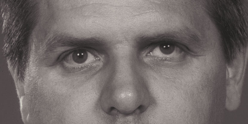
Does this person look normal to you?
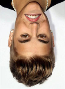
How about now?

The eyes, and the brain, can fall prey to a range of optical illusions, for a variety of reasons.
One such illusion is known as the Thatcher Effect. Named for former British Prime Minister Margaret Thatcher, since it was a photo of her that was famously used to illustrate the illusion, shows that the brain cannot properly process a photo of a face which is upside-down.
It demonstrates this by modifying upside down faces, flipping the mouth and eyes right side up. Because of this, and how our minds have learned to recognize faces, the brain believes nothing is wrong...until the image is turned right side up. Only at that point does the brain finally realize that yes, something was indeed very wrong with the image.
The Thatcher Effect demonstrates the importance of the brain’s processing of visual information, and the fact that recognizing faces is something we learn to do as our vision develops.
Read More
Optical illusions work by exploiting the disparity between what the eyes see and how the brain perceives that visual input. In doing so, they demonstrate that our visual systems “edit” what we see without us even being aware of it, as it decides what is worth paying attention to.
Even before humans understood the visual system as we do today, they created and were fascinated by optical illusions. Even today, we don’t know enough to explain how some of these fascinating illusions work.
Below are a few of these eye-twisting, and brain-fooling, illusions.
Lilac Chaser
[embed]https://youtu.be/a4fw3bmJwDs[/embed]
Focus on the crosshairs in the center of the image. After around 10-20 seconds, the lilac colored dots will start fading away to gray, while the dot that had previously been hopping around the lilac chain becomes a green dot, rotating in a circle.
This illusion is called Troxler’s fading, or Troxler’s effect, and it was discovered by Swiss polymath Ignaz Paul Troxler in 1804. Troxler’s effect is a result of the ability of our visual neurons to turn off their awareness of things which aren’t changing, and to heighten their perception of things which are.
Since, in the animated image, the lilac dots are stationary while the empty spot moves, after a short processing period, our visual system transitions to focusing solely on the moving blank dot. Due to a second illusion at play, the moving blank dot turns green. That is due to the retinas of the eyes becoming oversaturated with the lilac color from the dots we saw moments ago.When the lilac color is removed, what you see is its complementary color (a light green) in its place. Essentially, the green color is the result of white light with the lilac color subtracted.
Gradient Illusion
 The bar in the center of the image above appears gradated, changing from light to dark gray in the opposite direction as the background gradient.
Have you guessed the illusion yet? Perhaps you have. This is a case of your brain fooling itself. If you cover everything in the image apart from the bar, you’ll confirm that it is actually monochrome.
This illusion is caused by the brain interpreting the ends of the bar as being in different lighting, and from that determines what it thinks the bar would look like if evenly lit. Through this process, it decides that the left end of the bar is a light gray object in dim lighting, while the right is a darker gray that is well lit.
The bar in the center of the image above appears gradated, changing from light to dark gray in the opposite direction as the background gradient.
Have you guessed the illusion yet? Perhaps you have. This is a case of your brain fooling itself. If you cover everything in the image apart from the bar, you’ll confirm that it is actually monochrome.
This illusion is caused by the brain interpreting the ends of the bar as being in different lighting, and from that determines what it thinks the bar would look like if evenly lit. Through this process, it decides that the left end of the bar is a light gray object in dim lighting, while the right is a darker gray that is well lit.
Disappearing Dots
[embed]https://youtu.be/KK1hg5-xUm8[/embed]
For this illusion, start by staring at the blinking green light in the center of the video for several seconds. You’ll notice that as you continue staring, the yellow dots around the light. One of them might disappear, only to reappear moments later, or first one fades, then the other two, only for all 3 to come back after another few moments. As long as you keep focusing on the blinking light, the yellow dots continually disappear and reappear at random.
This illusion is called motion induced blindness, and it was first discovered in 1965, but it wasn’t until 2001, when it was rediscovered and named, that it gained significant attention. While similar to Troxler’s fading, which we touched on above, it is considered a separate phenomenon due to the requirement of a moving background for it to work.
There is still debate regarding the cause of motion induced blindness, with at least 5 theories being proposed.
One recent theory, known as the perceptual scotoma, proposes that this illusion is another attempt by the brain to provide us with clear and accurate perception. Since the 3 dots don’t change as the background spins, the visual system takes them out of our awareness, since they seem to be in contradiction with how the visual system understands things to work in real life. So, it essentially treats those spots like blind spots in the visual field.
The Hering Illusion

(Image credit: Fibonacci | Creative Commons)
This mind-bending illusion, first discovered in 1861 by German physiologist Ewald Hering, makes it look like the two straight red lines in fact bow outward. According to Hering, the reason for this illusion is that when we see the red lines crossing over the blue ones that radiate outward, our brains overestimate the angle made at the points where the red lines intersect the radiating ones.
Current research hypothesizes that the reason for this miscalculation is that since there is a delay between the time light hits the retina to when the brain perceives it, we evolved to compensate by generating images of what we think will occur one-tenth of a second in the future.
So in this case, that predicting leads to the overestimation of the angles, which makes the red lines look bent when they are truly straight. (And if you don’t believe it, cover up the blue lines and take another look at either of the red ones!)
The Checker Shadow Illusion
 On this checkerboard, the squares labeled A & B appear to be very different shades of gray, right? However, when we look at the board again, this time with solid gray lines running along both squares, we can see that they were the same shade all along!
This illusion, published by Edward H. Adelson, Professor of Vision Science at MIT in 1995, demonstrates how our visual system deals with shadows. When we try to determine the precise color of something, our brain knows that shadows can be misleading, in that they can make a surface look darker than it truly is. So the brain compensates by interpreting the shadowed surfaces as being lighter, even though it’s the exact same color as an unshadowed object which appears, at first glance, to be much darker.
On this checkerboard, the squares labeled A & B appear to be very different shades of gray, right? However, when we look at the board again, this time with solid gray lines running along both squares, we can see that they were the same shade all along!
This illusion, published by Edward H. Adelson, Professor of Vision Science at MIT in 1995, demonstrates how our visual system deals with shadows. When we try to determine the precise color of something, our brain knows that shadows can be misleading, in that they can make a surface look darker than it truly is. So the brain compensates by interpreting the shadowed surfaces as being lighter, even though it’s the exact same color as an unshadowed object which appears, at first glance, to be much darker.
Illusory Motion

(Image credit: Akiyoshi Kitaoka)
As hard as it might be to believe, nothing in this image is moving.
Unfortunately, there isn’t too much more we can tell you about this mind-boggling illusion.
To this day, there is no solid explanation for illusory motion. Some experts think it is linked to something called fixation jitter, involuntary eye movements which present the illusion that objects close to something you’re fixated on are moving.
Others believe that as you glance around the image, the motion detectors in your visual cortex get “confused” by dynamical changes in neurons, and thus believe that you are, in fact, seeing movement.
Illusions like this one serve to illustrate to us that while we know much, much more about vision than we did in the past, there is always more to learn!
These are just a few of the many fascinating optical illusions out there; demonstrations of just how fascinating our visual systems truly are.
Read More